日志
衰老(Senescence)
|
衰老: 从生物学上讲,衰老是生物随着时间的推移,自发的必然过程,它是复杂的自然现象,表现为结构和机能衰退,适应性和抵抗力减退。
http://baike.baidu.com/view/64548.htm
衰老的定义
衰老的概念
衰老(senility)是一种自然规律,因此,我们不可能违背这个规律。
但是,当人们采用良好的生活习惯和保健措施并适当地运动,就可以有效地延缓衰老,降低衰老相关疾病的发病率,提高生活质量。
衰老的实质与结果
衰老理论
机体变化
衰老的比较研究
衰老理论和原因
(一)体细胞突变学说
(二)自由基学说
(四)衰老的免疫学说
(五)端粒学说
Senescence http://www.biologyguide.net/human/senescence.htm
The Decline of Physiological Effectiveness- Rate of cell division and number of cells reduce
- All cells are capable to divide during embryological development
- Cells lose ability to divide after birth or have a lower growth rate
- Born with a fixed number of neurones → cannot divide/be replaced
- Decline in functional effectiveness of cells and organ systems
- Deterioration in cells / slower responds to stimuli / slows homeostatic mechanism / increases change of dysfunction and death
- Ageing is controlled by genes but can be slowed down by
- Regular (and adequate) sleep, (well balanced) meals, exercise
- Refrain from smoking and alcohol
- Keep body mass close to desirable mass for your height
- BMR
- Number of cells decreases during ageing → lowers BMR
- BMR decreases by ≈ 5% every 10 years above the age of 55
- 10-20 years - rapid decrease associated with adolescent growth spurt
- 20-35 - no change as body same size / same level of activity
- 30-70 - slow decrease associated with loss of muscles / gain of fat / reduced activity
- CARDIAC OUTPUT = STROKE VOLUME x HEART RATE
- Cardiac output decreases even though heart rate does not decline
- Due to cardiac muscle fibres weaken (mainly left ventricle)
- Decreases stroke volume of ventricles/volume of blood pumped per beat/cycle
- NERVE CONDUCTION VELOCITY
- Cells in peripheral nervous system and brain get less
- Neurones (nerve cells) are lost and cannot divide
- Effect of cell loss depends on cells location
- Brain loses ≈25% of cells that control muscular movement but hardly any that control speech → changes muscle coordination but not ability to speak
- LOSS OF MYELIN: no saltatory conduction / impulses cannot jump from node to node / impulses must pass through greater amount of membrane
- INCREASED WIDTH OF SYNAPSES: longer needed for diffusion/movement/greater distance to receptors/further to stimulate post-synaptic membrane/further diffusion distance of transmitter (across synapse)
- SLOWER SYNAPTIC TRANSMISSION: presynaptic neurones produce less neurotransmitter
- Cells in peripheral nervous system and brain get less
- Female reproductive capacity → MENOPAUSE (45-55 year old women)
- Ovaries gradually become insensitive to FSH / secretion of oestrogen becomes less / ovulation becomes less / menstrual cycle becomes less / vagina walls become thinner / woman is infertile when oestrogen secretion stops
- Levels of gonadotrophins (FSH, LH) rise to a peak after menopause
- At menopause, oestrogen no longer secreted
- FSH and LH no longer inhibited by negative feedback
- SYMPTOMS: due to loss of oestrogen
- Intense sweating / uncomfortable warmth / psychological problems
- Increase risk of osteoporosis (loss of bone tissue) and heart diseases
- TREATMENT: Hormone replacement therapy (HRT)
- Postmenstrual woman take in small doses of oestrogen and progesterone
- As tablets (orally) or apply implants beneath skin (skin patches)
Senescence (from Latin: senescere, meaning “to grow old,” from senex) or biological aging is the endogenous and hereditary process of accumulative changes to molecular and cellular structure disrupting metabolism with the passage of time, resulting in deterioration and death. Senescence occurs both on the level of the whole organism (organismal senescence) as well as on the level of its individual cells (cellular senescence). The science of biological aging is biogerontology. Albeit indirectly, senescence is by far the leading cause of death. Of the roughly 150,000 people who die each day across the globe, about two thirds—100,000 per day—die of age-related causes; in industrialized nations, moreover, the proportion is much higher, reaching 90%.[1] Senescence is not the inevitable fate of all organisms, and animal organisms of some groups (taxa) even experience chronological decrease in mortality, for all or part of their life cycle.[2] On the other extreme are accelerated aging diseases, rare in humans. There are a number of hypotheses as to why senescence occurs; for example, some posit it is programmed by gene expression changes, others that it is the cumulative damage caused by biological processes. Whether senescence as a biological process itself can be slowed down, halted or even reversed, is a subject of current scientific speculation and research.[3]
Contents [hide] |

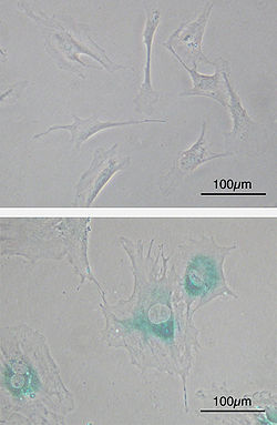
(upper) Primary mouse embryonic fibroblast cells (MEFs) before senescence. Spindle-shaped. (lower) MEFs became senescent after passages. Cells grow larger, flatten shape and expressed senescence-associated β-galactosidase (SABG, blue areas), a marker of cellular senescence.
Cellular senescence is the phenomenon by which normal diploid cells cease to divide, normally after about 50 cell divisions in vitro. This phenomenon is also known as "replicative senescence", the "Hayflick phenomenon", or the Hayflick limit in honour of Dr. Leonard Hayflick, co-author with Paul Moorhead, of the first paper describing it in 1961.[4] Cells can also be induced to senesce by certain toxins, irradiation, or the activation of certain oncogenes. In response to DNA damage (including shortened telomeres), cells either age or self-destruct (apoptosis, programmed cell death) if the damage cannot be easily repaired. In this 'cellular suicide', the death of one cell, or more, may benefit the organism as a whole. For example, in plants the death of the water-conducting xylem cells (tracheids and vessel elements) allows the cells to function more efficiently and so deliver water to the upper parts of a plant. The ones that do not self-destruct remain until destroyed by outside forces. Though they no longer replicate, senescent cells remain metabolically active and generally adopt phenotypes including flattened cell morphology, altered gene expression and secretion profiles (known as the senescence-associated secretory phenotype), and positive senescence-associated β-galactosidase staining.[5] In a study conducted in 2011 on mice, senescent cells were deliberately eradicated, which led to greater resistance against aging-associated diseases.[6] Cellular senescence is causally implicated in generating age-related phenotypes, and removal of senescent cells can prevent or delay tissue dysfunction and extend healthspan.[6]
Organismal senescence is the aging of whole organisms. In general, aging is characterized by the declining ability to respond to stress, increased homeostatic imbalance, and increased risk of aging-associated diseases. Death is the ultimate consequence of aging, though "old age" is not a scientifically recognized cause of death because there is always a specific proximal cause, such as cancer, heart disease, or liver failure. Aging of whole organisms is therefore a complex process that can be defined as "a progressive deterioration of physiological function, an intrinsic age-related process of loss of viability and increase in vulnerability".[7]
Differences in maximum life span among species correspond to different "rates of aging". For example, inherited differences in the rate of aging make a mouse elderly at 3 years and a human elderly at 80 years.[8] These genetic differences affect a variety of physiological processes, including the efficiency of DNA repair, antioxidant enzymes, and rates of free radical production.

Senescence of the organism gives rise to the Gompertz–Makeham law of mortality, which says that mortality rate accelerates rapidly with age.
Some animals, such as some reptiles and fish, age slowly (negligible senescence) and exhibit very long lifespans. Some even exhibit "negative senescence", in which mortality falls with age, in disagreement with the Gompertz–Makeham "law".[2]
Whether replicative senescence (Hayflick limit) plays a causative role in organismal aging is at present an active area of investigation.
The exact etiology of senescence is still largely unclear and yet to be discovered. The process of senescence is complex, and may derive from a variety of different mechanisms and exist for a variety of different reasons. However, senescence is not universal, and scientific evidence suggests that cellular senescence evolved in certain species because it prevents the onset of cancer. In a few simple species, such as those in the genus Hydra, senescence is negligible and cannot be detected.
All such species have no "post-mitotic" cells; they reduce the effect of damaging free radicals by cell division and dilution.[citation needed] Another related mechanism is that of the biologically immortal planarian flatworms, which have “apparently limitless [telomere] regenerative capacity fueled by a population of highly proliferative adult stem cells.”[9] These organisms are biologically immortal but not immortal in the traditional sense as they are nonetheless susceptible to trauma and infectious and non-infectious disease. Moreover, average lifespans can vary greatly within and between species. This suggests that both genetic and environmental factors contribute to aging. It has been said,[by whom?] for example, that exposure to ultraviolet light elevates accumulation of free-radical damage.
In general, theories that explain senescence have been divided between the programmed and stochastic theories of aging. Programmed theories imply that aging is regulated by biological clocks operating throughout the lifespan. This regulation would depend on changes in gene expression that affect the systems responsible for maintenance, repair, and defense responses. The Reproductive-Cell Cycle Theory suggests that aging is caused by changes in hormonal signaling over the lifespan.[10] Stochastic theories blame environmental impacts on living organisms that induce cumulative damage at various levels as the cause of aging, examples of which ranging from damage to DNA, damage to tissues and cells by oxygen radicals (widely known as free radicals countered by the even more well-known antioxidants), and cross-linking.
However, aging is seen as a progressive failure of homeodynamics (homeostasis) involving genes for the maintenance and repair, stochastic events leading to molecular damage and molecular heterogeneity, and chance events determining the probability of death. Since complex and interacting systems of maintenance and repair comprise the homeodynamic (old term: homeostasis) space of a biological system, aging is considered to be a progressive shrinkage of homeodynamic space mainly due to increased molecular heterogeneity.[citation needed]
A gene can be expressed at various stages of life. Therefore, natural selection can support lethal and harmful alleles, if their expression occurs after reproduction. Senescence may be the product of such selection.[11][12][13] In addition, aging is believed to have evolved because of the increasingly smaller probability of an organism still being alive at older age, due to predation and accidents, both of which may be random and age-invariant. It is thought[by whom?] that strategies that result in a higher reproductive rate at a young age, but shorter overall lifespan, result in a higher lifetime reproductive success and are therefore favoured by natural selection. In essence, aging is, therefore, the result of investing resources in reproduction, rather than maintenance of the body (the "Disposable Soma" theory[14]), in light of the fact that accidents, predation, and disease kill organisms regardless of how much energy is devoted to repair of the body. Various other theories of aging exist, and are not necessarily mutually exclusive.
The geneticist J. B. S. Haldane wondered why the dominant mutation that causes Huntington's disease remained in the population, and why natural selection had not eliminated it. The onset of this neurological disease is (on average) at age 45 and is invariably fatal within 10–20 years. Haldane assumed that, in human prehistory, few survived until age 45. Since few were alive at older ages and their contribution to the next generation was therefore small relative to the large cohorts of younger age groups, the force of selection against such late-acting deleterious mutations was correspondingly small. However, if a mutation affected younger individuals, selection against it would be strong. Therefore, late-acting deleterious mutations could accumulate in populations over evolutionary time through genetic drift. This principle has been demonstrated experimentally.[citation needed] And it is these later-acting deleterious mutations that are believed to allow—even cause—age-related mortality.
Peter Medawar formalised this observation in his mutation accumulation theory of aging.[15][16] "The force of natural selection weakens with increasing age—even in a theoretically immortal population, provided only that it is exposed to real hazards of mortality. If a genetic disaster… happens late enough in individual life, its consequences may be completely unimportant". The 'real hazards of mortality' are, in typical circumstances, predation, disease, and accidents. So, even an immortal population, whose fertility does not decline with time, will have fewer individuals alive in older age groups. This is called 'extrinsic mortality'. Young cohorts, not depleted in numbers yet by extrinsic mortality, contribute far more to the next generation than the few remaining older cohorts, so the force of selection against late-acting deleterious mutations, which affect only these few older individuals, is very weak. The mutations may not be selected against, therefore, and may spread over evolutionary time into the population.
The major testable prediction made by this model is that species that have high extrinsic mortality in nature will age more quickly and have shorter intrinsic lifespans. This is borne out among mammals, the best-studied in terms of life history. There is a correlation among mammals between body size and lifespan, such that larger species live longer than smaller species under controlled/optimum conditions, but there are notable exceptions. For instance, many bats and rodents are of similar size, yet bats live much longer. For instance, the little brown bat, half the size of a mouse, can live 30 years in the wild. A mouse will only live 2–3 years even under optimum conditions. The explanation is that bats have fewer predators, and therefore low extrinsic mortality. More individuals survive to later ages, so the force of selection against late-acting deleterious mutations is stronger. Fewer late-acting deleterious mutations equates to slower aging and therefore a longer lifespan. Birds are also warm-blooded and are similar in size to many small mammals, yet often live 5–10 times as long. They have less predation pressure than ground-dwelling mammals. Seabirds, which, in general, have the fewest predators of all birds, live longest.
When examining the body-size vs. lifespan relationship, one also observes that predatory mammals tend to live longer than prey mammals in a controlled environment, such as a zoo or nature reserve. The explanation for the long lifespans of primates (such as humans, monkeys, and apes) relative to body size is that their intelligence, and often their sociality, help them avoid becoming prey. High position in the food chain, intelligence and cooperativeness all reduce extrinsic mortality in species.
Another evolutionary theory of aging was proposed by George C. Williams[17] and involves antagonistic pleiotropy. A single gene may affect multiple traits. Some traits that increase fitness early in life may also have negative effects later in life. But, because many more individuals are alive at young ages than at old ages, even small positive effects early can be strongly selected for, and large negative effects later may be very weakly selected against. Williams suggested the following example: Perhaps a gene codes for calcium deposition in bones, which promotes juvenile survival and will therefore be favored by natural selection; however, this same gene promotes calcium deposition in the arteries, causing negative effects in old age. Thus, harmful biological changes in old age may result from selection for pleiotropic genes that are beneficial early in life but harmful later on. In this case, fitness is relatively high when Fisher's reproductive value is high and relatively low when Fisher's reproductive value is low.
| This section needs additional citations for verification. Please help improve this article by adding citations to reliable sources. Unsourced material may be challenged and removed. (July 2008) |
A number of genetic components of aging have been identified using model organisms, ranging from the simple budding yeast Saccharomyces cerevisiae to worms such as Caenorhabditis elegans and fruit flies (Drosophila melanogaster). Study of these organisms has revealed the presence of at least two conserved aging pathways.
One of these pathways involves the gene Sir2, a NAD+-dependent histone deacetylase. In yeast, Sir2 is required for genomic silencing at three loci: The yeast mating loci, the telomeres and the ribosomal DNA (rDNA). In some species of yeast, replicative aging may be partially caused by homologous recombination between rDNA repeats; excision of rDNA repeats results in the formation of extrachromosomal rDNA circles (ERCs). These ERCs replicate and preferentially segregate to the mother cell during cell division, and are believed to result in cellular senescence by titrating away (competing for) essential nuclear factors. ERCs have not been observed in other species (nor even all strains of the same yeast species) of yeast (which also display replicative senescence), and ERCs are not believed to contribute to aging in higher organisms such as humans (they have not been shown to accumulate in mammals in a similar manner to yeast). Extrachromosomal circular DNA (eccDNA) has been found in worms, flies, and humans. The origin and role of eccDNA in aging, if any, is unknown.
Despite the lack of a connection between circular DNA and aging in higher organisms, extra copies of Sir2 are capable of extending the lifespan of both worms and flies (though, in flies, this finding has not been replicated by other investigators, and the activator of Sir2 resveratrol does not reproducibly increase lifespan in either species[18]). Whether the Sir2 homologues in higher organisms have any role in lifespan is unclear, but the human SIRT1 protein has been demonstrated to deacetylate p53, Ku70, and the forkhead family of transcription factors. SIRT1 can also regulate acetylates such as CBP/p300, and has been shown to deacetylate specific histone residues.
RAS1 and RAS2 also affect aging in yeast and have a human homologue. RAS2 overexpression has been shown to extend lifespan in yeast.
Other genes regulate aging in yeast by increasing the resistance to oxidative stress. Superoxide dismutase, a protein that protects against the effects of mitochondrial free radicals, can extend yeast lifespan in stationary phase when overexpressed.
In higher organisms, aging is likely to be regulated in part through the insulin/IGF-1 pathway. Mutations that affect insulin-like signaling in worms, flies, and the growth hormone/IGF1 axis in mice are associated with extended lifespan. In yeast, Sir2 activity is regulated by the nicotinamidase PNC1. PNC1 is transcriptionally upregulated under stressful conditions such as caloric restriction, heat shock, and osmotic shock. By converting nicotinamide to niacin, nicotinamide is removed, inhibiting the activity of Sir2. A nicotinamidase found in humans, known as PBEF, may serve a similar function, and a secreted form of PBEF known as visfatin may help to regulate serum insulin levels. It is not known, however, whether these mechanisms also exist in humans, since there are obvious differences in biology between humans and model organisms.
Sir2 activity has been shown to increase under calorie restriction. Due to the lack of available glucose in the cells, more NAD+ is available and can activate Sir2. Resveratrol, a stilbenoid found in the skin of red grapes, was reported to extend the lifespan of yeast, worms, and flies (the lifespan extension in flies and worms have proved to be irreproducible by independent investigators[18]). It has been shown to activate Sir2 and therefore mimics the effects of calorie restriction, if one accepts that caloric restriction is indeed dependent on Sir2.
Gene expression is imperfectly controlled, and it is possible that random fluctuations in the expression levels of many genes contribute to the aging process as suggested by a study of such genes in yeast.[19] Individual cells, which are genetically identical, none-the-less can have substantially different responses to outside stimuli, and markedly different lifespans, indicating the epigenetic factors play an important role in gene expression and aging as well as genetic factors.
According to the GenAge database of aging-related genes there are over 700 genes associated with aging in model organisms: 555 in the soil roundworm (Caenorhabditis elegans), 87 in the bakers' yeast (Saccharomyces cerevisiae), 75 in the fruit fly (Drosophila melanogaster) and 68 in the mouse (Mus musculus).[20] The following is a list of genes connected to longevity through research[citation needed] on model organisms:
| Podospora | Saccharomyces | Caenorhabditis | Drosophila | Mus |
|---|---|---|---|---|
| grisea | LAG1 | daf-2 | sod1 | Prop-1 |
| LAC1 | age-1/daf-23 | cat1 | p66shc (Not independently verified) | |
| pit-1 | Ghr | |||
| RAS1 | daf-18 | mth | mclk1 | |
| RAS2 | akt-1/akt-2 | |||
| PHB1 | daf-16 | |||
| PHB2 | daf-12 | |||
| CDC7 | ctl-1 | |||
| BUD1 | old-1 | |||
| RTG2 | spe-26 | |||
| RPD3 | clk-1 | |||
| HDA1 | mev-1 | |||
| SIR2 | ||||
| aak-2 | ||||
| SIR4-42 | ||||
| UTH4 | ||||
| YGL023 | ||||
| SGS1 | ||||
| RAD52 | ||||
| FOB1 |
As noted above, senescence is not universal. It was once thought that senescence did not occur in single-celled organisms that reproduce through the process of cellular mitosis.[21] Recent investigation has unveiled a more complex picture. Single cells do accumulate age-related damage. On mitosis the debris is not evenly divided between the new cells. Instead it passes to one of the cells leaving the other cell pristine. With successive generations the cell population becomes a mosaic of cells with half ageless and the rest with varying degrees of senescence.[22]
Moreover, cellular senescence is not observed in several organisms, including perennial plants, sponges, corals, and lobsters. In those species where cellular senescence is observed, cells eventually become post-mitotic when they can no longer replicate themselves through the process of cellular mitosis; i.e., cells experience replicative senescence. How and why some cells become post-mitotic in some species has been the subject of much research and speculation, but (as noted above) it is sometimes suggested that cellular senescence evolved as a way to prevent the onset and spread of cancer. Somatic cells that have divided many times will have accumulated DNA mutations and would therefore be in danger of becoming cancerous if cell division continued.
Lately, the role of telomeres in cellular senescence has aroused general interest, especially with a view to the possible genetically adverse effects of cloning. The successive shortening of the chromosomal telomeres with each cell cycle is also believed to limit the number of divisions of the cell, thus contributing to aging. There have, on the other hand, also been reports that cloning could alter the shortening of telomeres. Some cells do not age and are, therefore, described as being "biologically immortal". It is theorized by some that when it is discovered exactly what allows these cells, whether it be the result of telomere lengthening or not, to divide without limit that it will be possible to genetically alter other cells to have the same capability. It is further theorized that it will eventually be possible to genetically engineer all cells in the human body to have this capability by employing gene therapy and, therefore, stop or reverse aging, effectively making the entire organism potentially immortal.
The length of the telomere strand has senescent effects; telomere shortening activates extensive alterations in alternative RNA splicing that produce senescent toxins such as progerin, which degrades the tissue and makes it more prone to failure.[23]
Cancer cells are usually immortal. In about 85% of tumors, this evasion of cellular senescence is the result of up-activation of their telomerase genes.[24] This simple observation suggests that reactivation of telomerase in healthy individuals could greatly increase their cancer risk.
A research team led by Darren J. Baker, Tamara Tchkonia, James L. Kirkland, and Jan M. van Deursen at the Mayo Clinic in Rochester, Minn., purged all the senescent cells in mice by giving them a drug that forces the cells to self-destruct. The mice’s tissues showed a major improvement in the usual burden of age-related disorders. They did not develop cataracts, avoided the usual wasting of muscle with age, and could exercise much longer on a mouse treadmill. They retained the fat layers in the skin that usually thin out with age and, in people, cause wrinkling.[6]
One of the earliest aging theories was the Rate of Living Hypothesis described by Raymond Pearl in 1928[25] (based on earlier work by Max Rubner), which states that fast basal metabolic rate corresponds to short maximum life span.
While there may be some validity to the idea that for various types of specific damage detailed below that are by-products of metabolism, all other things being equal, a fast metabolism may reduce lifespan, in general this theory does not adequately explain the differences in lifespan either within, or between, species. Calorically-restricted animals process as much, or more, calories per gram of body mass, as their ad libitum fed counterparts, yet exhibit substantially longer lifespans.[citation needed] Similarly, metabolic rate is a poor predictor of lifespan for birds, bats and other species that, it is presumed, have reduced mortality from predation, and therefore have evolved long lifespans even in the presence of very high metabolic rates.[26] In a 2007 analysis it was shown that, when modern statistical methods for correcting for the effects of body size and phylogeny are employed, metabolic rate does not correlate with longevity in mammals or birds.[27] (For a critique of the Rate of Living Hypothesis see Living fast, dying when?[28])
With respect to specific types of chemical damage caused by metabolism, it is suggested that damage to long-lived biopolymers, such as structural proteins or DNA, caused by ubiquitous chemical agents in the body such as oxygen and sugars, are in part responsible for aging. The damage can include breakage of biopolymer chains, cross-linking of biopolymers, or chemical attachment of unnatural substituents (haptens) to biopolymers.
Under normal aerobic conditions, approximately 4% of the oxygen metabolized by mitochondria is converted to superoxide ion, which can subsequently be converted to hydrogen peroxide, hydroxyl radical and eventually other reactive species including other peroxides and singlet oxygen, which can, in turn, generate free radicals capable of damaging structural proteins and DNA. Certain metal ions found in the body, such as copper and iron, may participate in the process. (In Wilson's disease, a hereditary defect that causes the body to retain copper, some of the symptoms resemble accelerated senescence.) These processes termed oxidative stress are linked to the potential benefits of dietary polyphenol antioxidants, for example in coffee,[29] red wine and tea.[30]
Sugars such as glucose and fructose can react with certain amino acids such as lysine and arginine and certain DNA bases such as guanine to produce sugar adducts, in a process called glycation. These adducts can further rearrange to form reactive species, which can then cross-link the structural proteins or DNA to similar biopolymers or other biomolecules such as non-structural proteins. People with diabetes, who have elevated blood sugar, develop senescence-associated disorders much earlier than the general population, but can delay such disorders by rigorous control of their blood sugar levels. There is evidence that sugar damage is linked to oxidant damage in a process termed glycoxidation.
Free radicals can damage proteins, lipids or DNA. Glycation mainly damages proteins. Damaged proteins and lipids accumulate in lysosomes as lipofuscin. Chemical damage to structural proteins can lead to loss of function; for example, damage to collagen of blood vessel walls can lead to vessel-wall stiffness and, thus, hypertension, and vessel wall thickening and reactive tissue formation (atherosclerosis); similar processes in the kidney can lead to renal failure. Damage to enzymes reduces cellular functionality. Lipid peroxidation of the inner mitochondrial membrane reduces the electric potential and the ability to generate energy. It is probably no accident that nearly all of the so-called "accelerated aging diseases" are due to defective DNA repair enzymes.
It is believed that the impact of alcohol on aging can be partly explained by alcohol's activation of the HPA axis, which stimulates glucocorticoid secretion, long-term exposure to which produces symptoms of aging.[31]
Alexander[32] was the first to propose that DNA damage is the primary cause of aging. Early experimental evidence supporting this idea was reviewed by Gensler and Bernstein.[33] By the early 1990s experimental support for this proposal was substantial, and further indicated that DNA damage due to reactive oxygen species was a major source of the DNA damages causing aging.[34][35][36][37][38] The current state of evidence bearing on this theory is reviewed in DNA damage theory of aging by Bernstein et al.[39]
Reliability theory suggests that biological systems start their adult life with a high load of initial damage. Reliability theory is a general theory about systems failure. It allows researchers to predict the age-related failure kinetics for a system of given architecture (reliability structure) and given reliability of its components. Reliability theory predicts that even those systems that composed entirely of non-aging elements (with a constant failure rate) will nevertheless deteriorate (fail more often) with age, if these systems are redundant in irreplaceable elements. Aging, therefore, is a direct consequence of systems.
Reliability theory also predicts the late-life mortality deceleration with subsequent leveling-off, as well as the late-life mortality plateaus, as an inevitable consequence of redundancy exhaustion at extreme old ages. The theory explains why mortality rates increase exponentially with age (the Gompertz law) in many species, by taking into account the initial flaws (defects) in newly formed systems. It also explains why organisms "prefer" to die according to the Gompertz law, while technical devices usually fail according to the Weibull (power) law. Reliability theory allows to specify conditions when organisms die according to the Weibull distribution: Organisms should be relatively free of initial flaws and defects. The theory makes it possible to find a general failure law applicable to all adult and extreme old ages, where the Gompertz and the Weibull laws are just special cases of this more general failure law. The theory explains why relative differences in mortality rates of compared populations (within a given species) vanish with age (compensation law of mortality), and mortality convergence is observed due to the exhaustion of initial differences in redundancy levels.
A set of rare hereditary (genetic) disorders, each called progeria, has been known for some time. Sufferers exhibit symptoms resembling accelerated aging, including wrinkled skin. The cause of Hutchinson–Gilford progeria syndrome was reported in the journal Nature in May 2003.[40] This report suggests that DNA damage, not oxidative stress, is the cause of this form of accelerated aging.
Recently, a kind of early senescence has been alleged to be a possible unintended outcome of early cloning experiments. The issue was raised in the case of Dolly the sheep, following her death from a contagious lung disease. The claim that Dolly's early death involved premature senescence has been vigorously contested,[41] and Dolly's creator, Dr. Ian Wilmut has expressed the view that her illness and death were probably unrelated to the fact that she was a clone.
| Look up senescence in Wiktionary, the free dictionary. |
- ^ Aubrey D.N.J, de Grey (2007). "Life Span Extension Research and Public Debate: Societal Considerations" (PDF). Studies in Ethics, Law, and Technology 1 (1). doi:10.2202/1941-6008.1011. Article 5. http://www.sens.org/files/pdf/ENHANCE-PP.pdf.
- ^ a b Ainsworth, C; Lepage, M (2007). "Evolution's greatest mistakes". The New Scientist 195 (2616): 36–39. doi:10.1016/S0262-4079(07)62033-8.
- ^ "SENS Foundation". http://sens.org/.
- ^ Hayflick L, Moorhead PS (December 1961). "The serial cultivation of human diploid cell strains". Exp. Cell Res. 25: 585–621. PMID 13905658.
- ^ Campisi, Judith (February 2013). "Aging, Cellular Senescence, and Cancer". Annual Review of Physiology 75: 685–705. doi:10.1146/annurev-physiol-030212-183653. PMID 23140366.
- ^ a b c Baker, D.; Wijshake, T.; Tchkonia, T.; LeBrasseur, N.; Childs, B.; van de Sluis, B.; Kirkland, J.; van Deursen, J. (Nov 10 2011). "Clearance of p16Ink4a-positive senescent cells delays ageing-associated disorders". Nature 479: 232–6. doi:10.1038/nature10600.
- ^ "Aging and Gerontology Glossary". http://www.senescence.info/glossary.html. Retrieved 2011-02-26.
- ^ Austad, S (2009). "Comparative Biology of Aging". J Gerontol a Biol Sci Med Sci 64 (2): 199–201. doi:10.1093/gerona/gln060. PMC 2655036. PMID 19223603. //www.ncbi.nlm.nih.gov/pmc/articles/PMC2655036/.
- ^ Thomas C. J. Tan, Ruman Rahman, Farah Jaber-Hijazi, Daniel A. Felix, Chen Chen, Edward J. Louis, and Aziz Aboobaker (February 2012). "Telomere maintenance and telomerase activity are differentially regulated in asexual and sexual worms". PNAS 109 (9). doi:10.1073/pnas.1118885109. http://www.pnas.org/content/early/2012/02/22/1118885109.full.pdf+html.
- ^ Bowen RL, Atwood CS (2011). "The reproductive-cell cycle theory of aging: an update.". Experimental Gerontology 46 (2): 100–7. doi:10.1016/j.exger.2010.09.007. PMID 20851172.
- ^ Medawar, P.B. (1952). An Unsolved problem of biology; an inaugural lecture delivered at University College, London, 6 December, 1951. London: H.K. Lewis. OCLC 8482093.
- ^ Williams, G.C. (1957). "Pleiotropy, Natural Selection, and the Evolution of Senescence". Evolution 11: 398-411.
- ^ Hamilton WD (September 1966). "The moulding of senescence by natural selection". J. Theor. Biol. 12 (1): 12–45. PMID 6015424. http://linkinghub.elsevier.com/retrieve/pii/0022-5193(66)90184-6.
- ^ Kirkwood TB (November 1977). "Evolution of ageing". Nature 270 (5635): 301–4. doi:10.1038/270301a0. PMID 593350.
- ^ Medawar PB (1946). "Old age and natural death". Modern Quarterly 1: 30–56.
- ^ Medawar, Peter B. (1952). An Unsolved Problem of Biology. London: H. K. Lewis.[page needed]
- ^ Williams, George C. (December 1957). "Pleiotropy, Natural Selection, and the Evolution of Senescence". Evolution 11 (4): 398–411. doi:10.2307/2406060. JSTOR 2406060. http://www.devonescent.org/pdfs/Williams57.pdf.
- ^ a b Bass TM, Weinkove D, Houthoofd K, Gems D, Partridge L (October 2007). "Effects of resveratrol on lifespan in Drosophila melanogaster and Caenorhabditis elegans". Mechanisms of Ageing and Development 128 (10): 546–52. doi:10.1016/j.mad.2007.07.007. PMID 17875315.
- ^ Ryley J, Pereira-Smith OM (2006). "Microfluidics device for single cell gene expression analysis in Saccharomyces cerevisiae". Yeast 23 (14–15): 1065–73. doi:10.1002/yea.1412. PMID 17083143.
- ^ "GenAge database". http://genomics.senescence.info/genes/models.html. Retrieved 2011-02-26.
- ^ Gavrilov LA, Gavrilova NS (December 2001). "The reliability theory of aging and longevity". Journal of Theoretical Biology 213 (4): 527–45. doi:10.1006/jtbi.2001.2430. PMID 11742523.
- ^ Stephens C (April 2005). "Senescence: even bacteria get old". Curr. Biol. 15 (8): R308–10. doi:10.1016/j.cub.2005.04.006. PMID 15854899. http://linkinghub.elsevier.com/retrieve/pii/S0960-9822(05)00377-5.
- ^ Collins FS et al. (Jun 13 2011). "Progerin and telomere dysfunction collaborate to trigger cellular senescence in normal human fibroblasts". J Clin Invest. 121 (7): 2833–44. doi:10.1172/JCI43578. PMC 3223819. PMID 21670498. //www.ncbi.nlm.nih.gov/pmc/articles/PMC3223819/.
- ^ Hanahan D, Weinberg RA (January 2000). "The hallmarks of cancer". Cell 100 (1): 57–70. doi:10.1016/S0092-8674(00)81683-9. PMID 10647931.
- ^ Pearl, Raymond (1928). The Rate of Living, Being an Account of Some Experimental Studies on the Biology of Life Duration. New York: Alfred A. Knopf.[page needed]
- ^ Brunet-Rossinni AK, Austad SN (2004). "Ageing studies on bats: a review". Biogerontology 5 (4): 211–22. doi:10.1023/B:BGEN.0000038022.65024.d8. PMID 15314271.
- ^ de Magalhães JP, Costa J, Church GM (1 February 2007). "An Analysis of the Relationship Between Metabolism, Developmental Schedules, and Longevity Using Phylogenetic Independent Contrasts". The Journals of Gerontology Series A: Biological Sciences and Medical Sciences 62 (2): 149–60. doi:10.1093/gerona/62.2.149. PMC 2288695. PMID 17339640. http://biomed.gerontologyjournals.org/cgi/pmidlookup?view=long&pmid=17339640.[dead link]
- ^ Speakman JR, Selman C, McLaren JS, Harper EJ (1 June 2002). "Living fast, dying when? The link between aging and energetics". The Journal of Nutrition 132 (6 Suppl 2): 1583S–97S. PMID 12042467. http://jn.nutrition.org/cgi/pmidlookup?view=long&pmid=12042467.
- ^ Freedman ND, Park Y, Abnet CC, Hollenbeck AR, Sinha R (May 2012). "Association of coffee drinking with total and cause-specific mortality". N. Engl. J. Med. 366 (20): 1891–904. doi:10.1056/NEJMoa1112010. PMC 3439152. PMID 22591295. http://www.nejm.org/doi/full/10.1056/NEJMoa1112010.
- ^ Yang Y, Chan SW, Hu M, Walden R, Tomlinson B (2011). "Effects of some common food constituents on cardiovascular disease". ISRN Cardiol 2011: 397136. doi:10.5402/2011/397136. PMC 3262529. PMID 22347642. //www.ncbi.nlm.nih.gov/pmc/articles/PMC3262529/.
- ^ Spencer RL, Hutchison KE (1999). "Alcohol, aging, and the stress response". Alcohol Research & Health 23 (4): 272–83. PMID 10890824. http://pubs.niaaa.nih.gov/publications/arh23-4/272-283.pdf.
- ^ Alexander P (1967). "The role of DNA lesions in the processes leading to aging in mice". Symp. Soc. Exp. Biol. 21: 29–50. PMID 4860956.
- ^ Gensler HL, Bernstein H (September 1981). "DNA damage as the primary cause of aging". Q Rev Biol 56 (3): 279–303. doi:10.1086/412317. PMID 7031747.
- ^ Bernstein C, Bernstein H (1991). Aging, Sex, and DNA Repair. San Diego CA: Academic Press. ISBN 0123960037.
- ^ Ames BN, Gold LS (1991). "Endogenous mutagens and the causes of aging and cancer". Mutat. Res. 250 (1-2): 3–16. PMID 1944345.
- ^ Holmes GE, Bernstein C, Bernstein H (September 1992). "Oxidative and other DNA damages as the basis of aging: a review". Mutat. Res. 275 (3-6): 305–15. doi:10.1016/0921-8734(92)90034-M. PMID 1383772.
- ^ Rao KS, Loeb LA (September 1992). "DNA damage and repair in brain: relationship to aging". Mutat. Res. 275 (3-6): 317–29. doi:10.1016/0921-8734(92)90035-N. PMID 1383773.
- ^ Ames BN, Shigenaga MK, Hagen TM (September 1993). "Oxidants, antioxidants, and the degenerative diseases of aging". Proc. Natl. Acad. Sci. U.S.A. 90 (17): 7915–22. doi:10.1073/pnas.90.17.7915. PMC 47258. PMID 8367443. http://www.pnas.org/cgi/pmidlookup?view=long&pmid=8367443.
- ^ Bernstein H, Payne CM, Bernstein C, Garewal H, Dvorak K (2008). "Ch. 1: Cancer and aging as consequences of un-repaired DNA damage". In Honoka Kimura, Aoi Suzuki. New Research on DNA Damages. New York: Nova Science. pp. 1–47. ISBN 1604565810.
- ^ Mounkes LC, Kozlov S (2003). "A progeroid syndrome in mice is caused by defects in A-type lamins". Nature 423 (6937): 298–301. http://www.nature.com.gate2.inist.fr/nature/journal/v423/n6937/pdf/nature01631.pdf.
- ^ Lynn Macintosh, Kerry (2005). Illegal Beings: Human Clones and the Law. Cambridge: Cambridge University Press. ISBN 0-521-85328-1.[page needed]
| Look up senescence in Wiktionary, the free dictionary. |
全部作者的其他最新日志
热门日志导读
- • 感受
- • 努力挣威望
- • “干细胞”滥用:救命还是敛财?
- • 慢慢体会的小故事
- • 细胞培养板的选择
- • miRNA-操纵干细胞命运


